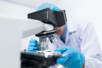

Science moves fast when researchers can actually see what is happening inside living systems. Many breakthroughs start with a clear picture. That is why fluorescence microscopy has become such a core part of modern research. It gives teams the ability to track tiny details that once stayed hidden. It turns complex biology into something visual and understandable. It also helps scientists move from guesswork to insight with more confidence and speed.
A Closer Look at Cell Behavior
Researchers often begin with basic cell studies. They want to see how cells grow, move, and react to changes. Fluorescence microscope images make that possible. They highlight proteins, pathways, and structures that stay invisible with standard light microscopy.
Scientists can follow single cells across time. They can track small changes in shape and function. They can observe how cells respond to stress or treatment. These images give a deeper sense of what is actually happening inside the sample. The detail helps teams ask better questions and build stronger experiments.
This kind of imaging also helps new researchers learn faster. They can watch living systems in real time. It makes the science feel more hands-on and more meaningful.
Mapping How Molecules Interact
Life science research often focuses on relationships between molecules. Small interactions can change the direction of a whole biological process. Fluorescence microscopy reveals those connections with clarity.
Scientists can label one molecule in green and another in red. When they overlap, the colors mix. This simple visual cue shows when and where molecules meet. It also shows how long they stay together and what happens next.
This helps teams understand protein networks. It helps them follow signaling pathways. It reveals how cells communicate or stop communicating. These insights often guide new therapies. They also support early-stage drug development by helping researchers see what works and what does not.
Tracking Disease Progression
Disease research relies on detail. Scientists must understand how illnesses start and how they spread. Fluorescence microscopy gives them the level of visibility they need.
Researchers can watch how viruses enter cells. They can see where bacteria gather or where cancer cells migrate. They can track markers that signal inflammation or tissue damage. Each image becomes a clue in a larger story.
This kind of tracking helps labs test treatments with accuracy. They can see if a therapy reaches the right place. They can watch how cells react after exposure. They can spot early signs of success or failure. It keeps projects moving forward with fewer setbacks.
Improving Precision in Drug Discovery
Drug development is one of the hardest parts of life science. It demands careful testing and layers of validation. Fluorescence microscopy gives researchers more control.
Teams can observe how compounds behave inside cells. They can see if a drug binds to the right target. They can watch for toxic side effects long before animal or human testing. This saves time and resources. It also makes early screening more reliable.
Scientists can also use fluorescence markers to check drug delivery systems. They can track how nanoparticles move through tissue. They can confirm whether a delivery method works as planned. These steps help labs refine drug candidates with more accuracy.
Supporting Breakthroughs in Genetics
Genetic research depends on visualization. DNA, RNA, and gene expression patterns tell important stories. Fluorescence microscopy helps researchers see those stories with greater clarity.
Teams can label specific genes. They can measure expression levels at different stages. They can observe how changes in the genome affect cell behavior. These images show the direct impact of genetic edits or mutations. They also reveal new possibilities for gene-based therapies.
Fluorescence microscopy supports many techniques in genomic research. It guides CRISPR work. It verifies gene delivery. It helps confirm that edits happened in the right place. These steps boost trust in the final results.
Bringing New Power to Tissue Engineering
Tissue engineering requires constant observation. Cells must grow into structures that mimic real tissues. Fluorescence microscopy helps researchers check these structures with precision.
Scientists can see how cells attach to scaffolds. They can measure how fast they spread. They can watch the formation of blood vessel networks. They can test how tissues respond to pressure or chemical signals. These images show progress that researchers would never notice with the naked eye.
This type of imaging also helps teams evaluate their materials. They can check if a scaffold supports healthy cell growth. They can monitor nutrient flow. They can detect early stress signals in engineered tissues. These details support safer and more reliable development.
A Tool That Pushes Research Forward
Fluorescence microscopy continues to grow in importance. Each year brings new dyes, new lenses, and new imaging techniques. These upgrades give researchers sharper images and more control. They also open new areas of study that once felt impossible.
The value of fluorescence microscopy does not come from the tool alone. It comes from the people who use it. Skilled researchers can turn images into answers. They can turn patterns into hypotheses. They can turn ideas into discoveries. That is why fluorescence microscopy sits at the center of so many major breakthroughs in life science.
Final Thoughts
Fluorescence microscopy gives researchers the ability to see more, understand more, and move faster. It helps teams explore cell behavior. It supports disease studies. It pushes drug discovery forward. It strengthens genetic research. It guides tissue engineering. It sits quietly in the background while it transforms science at every level.
Labs that rely on these imaging methods move with better accuracy and greater confidence. They can test ideas sooner. They can refine projects with fewer delays. They can reach insights that shape the future of life science.